MoleMax HD High-Definition Digital Dermoscopy System for Clinics
Digital Skin Imaging System
MoleMax HD is a modular, subscription‑free digital dermoscopy platform delivering ultra‑high resolution imaging (up to 100x magnification) with polarised and non‑polarised modes. Designed for dermatologists and skin‑cancer GPs, MoleMax HD improves diagnostic accuracy, streamlines workflow and enhances patient engagement.
Key features
Clinical benefits

Molemax Systems
Digital Skin Imaging System
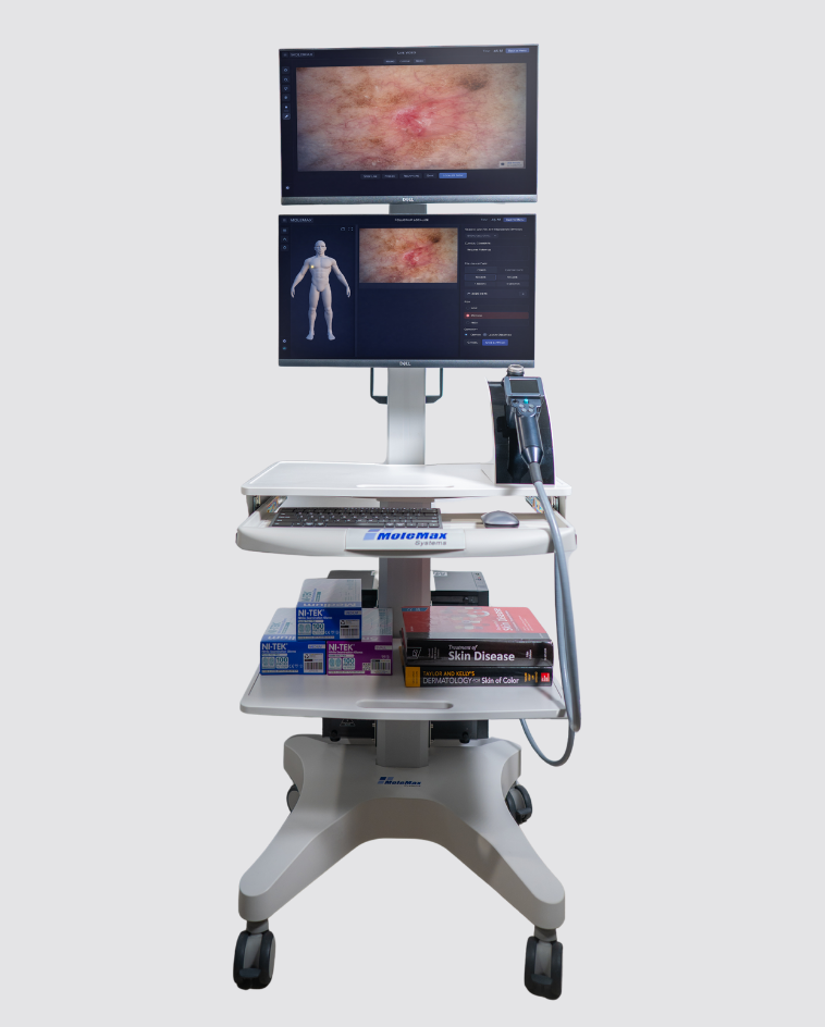
The MoleMax HD Trolley pairs the HD imaging platform with a lightweight, height‑adjustable ergonomic trolley for flexible room‑to‑room use. It provides a customizable workstation with dual touch monitors, camera dock and cable management to speed consultations and improve patient education.
Key features
Clinical benefits

Molemax Systems
MoleMax HD software is built for clinical efficiency: total body mapping, real‑time overlay follow‑up, lesion scoring (Expertizer), and an extensive skin reference library — all without recurring subscription fees. It supports patient management, PDF reporting and longitudinal comparisons. (Source: brochure pages 8, 12)


*An optional extra. Additional charges apply.

*An optional extra. Additional charges apply.


*An optional extra. Additional charges apply.


MoleMax Systems
Our Practice Management Link will improve the workflow, cost efficiency and time management of your practice, linking the MoleMax software to your Practice Management Software and integrate with your patient notes.
Digital Skin Imaging System
MoleMax HD is a modular, subscription‑free digital dermoscopy platform delivering ultra‑high resolution imaging (up to 100x magnification) with polarised and non‑polarised modes. Designed for dermatologists and skin‑cancer GPs, MoleMax HD improves diagnostic accuracy, streamlines workflow and enhances patient engagement.
Key features
Clinical benefits
Molemax Systems
Digital Skin Imaging System
The MoleMax HD Trolley pairs the HD imaging platform with a lightweight, height‑adjustable ergonomic trolley for flexible room‑to‑room use. It provides a customizable workstation with dual touch monitors, camera dock and cable management to speed consultations and improve patient education.
Key features
Clinical benefits
Molemax Systems
MoleMax HD software is built for clinical efficiency: total body mapping, real‑time overlay follow‑up, lesion scoring (Expertizer), and an extensive skin reference library — all without recurring subscription fees. It supports patient management, PDF reporting and longitudinal comparisons. (Source: brochure pages 8, 12)
*An optional extra. Additional charges apply.
*An optional extra. Additional charges apply.
*An optional extra. Additional charges apply.
MoleMax Systems
Our Practice Management Link will improve the workflow, cost efficiency and time management of your practice, linking the MoleMax software to your Practice Management Software and integrate with your patient notes.
MoleMax Systems, a division of Macquarie Medical Systems, specialises in the development and production of dermatoscopic hardware and software, including dermatoscopes and the MoleMax Dermoscopy Software. These integrated solutions are designed for early clinical imaging sessions, analysis and comparisons of skin cancer, providing dermatologists with advanced tools for high-resolution imaging and analysis of skin lesions to improve patient outcomes.
Subscribe To The MoleMax Newsletter