Stethoscope, otoscope, ophthalmoscope. Essential diagnostic tools that no primary health care practitioner can manage without. In this decade, another should be added to the list – the dermoscope.
A Brief History of Dermoscopy.
The first handheld dermatoscopes became available in the late 1980s and over the ensuing 2 decades both the instruments themselves and the science underpinning their use were refined, initially for pigmented lesions and later for non-pigmented lesions. Training courses, both face-to-face and virtual, were developed so that dermatoscope users could understand and interpret the colours and structures they were seeing. Today, there is overwhelming evidence that dermoscopy significantly improves the accuracy of diagnosis of skin lesions.
What is Dermoscopy?
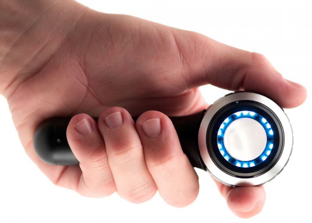
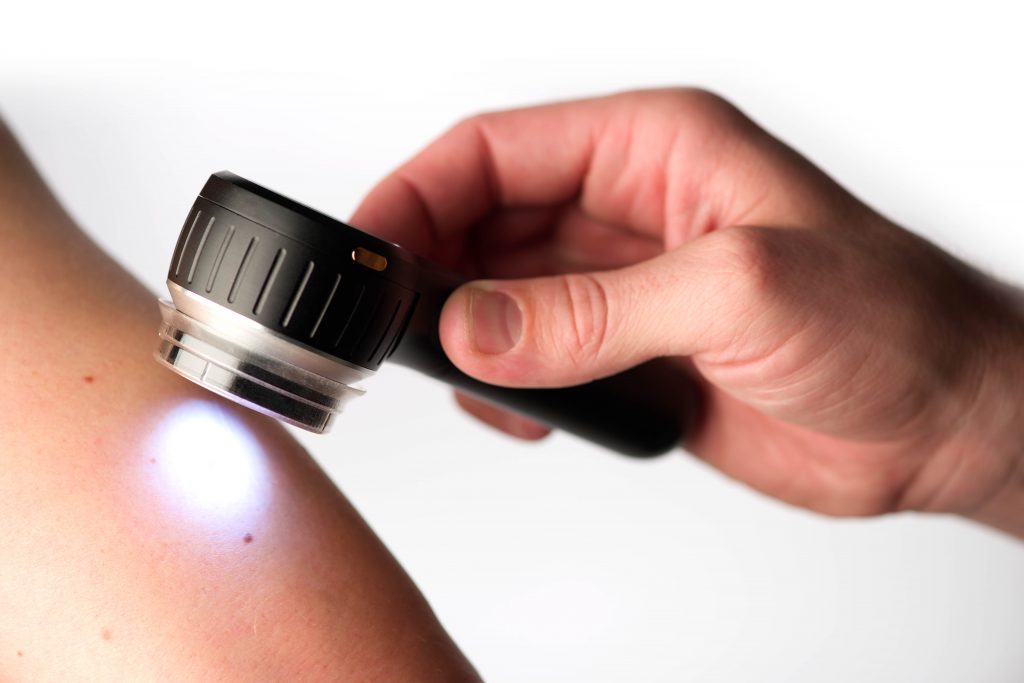
A non-invasive method to view and evaluate in vivo the colours and microstructures of epidermis, dermo-epidermal junction and the papillary dermis, which are not visible to the naked eye. The dermatoscope eliminates surface reflection making the stratum corneum transparent and allowing examination of the epidermis and superficial dermis. Most dermatoscopes provide x10 magnification.
Why Has the Dermatoscope Become Essential?
Australasia has the highest incidence of skin cancer in the world. The sheer volume of disease means that the diagnosis and management of most skin cancer is carried out in primary care with only the more difficult, sinister or advanced tumours being referred into secondary care. A high level of diagnostic accuracy amongst GPs is essential in order to avoid missing melanomas and at the same time to reduce unnecessary excisions of benign lesions. A recently published document, Quality Statements to Guide Melanoma Diagnosis and Treatment in New Zealand, includes the following.
All primary health care professionals involved in early detection of melanoma should be trained in the use of the dermatoscope and regularly undertake refresher training.
In primary health care practices, [patients should have] access to at least one designated primary health care professional trained in the dermatoscopic diagnosis and management of melanoma.
Training.
Medical students all receive training in the use of the scopes mentioned at the start of this article. Some universities now include some dermatoscopy training their curricula, but for those who trained in the previous millennium, there was none. In the same way that dermatopathologists require training to be able to accurately diagnose what they see under the microscope, so do dermatoscopists. However, there is good evidence that, for the novice, a few hours training and the use of an algorithm significantly improves diagnostic accuracy.
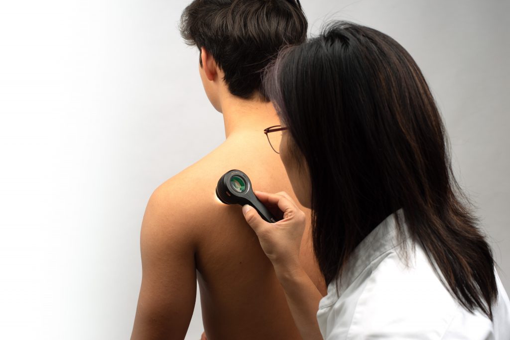
“Can you please have a look at this spot on my skin?”
As a GP, how many times have you been asked this at the end of a consultation? If you have a dermatoscope on your desk and have undertaken the training, you can examine the lesion and quite rapidly assign it to 1 of 4 categories.
- A confident, specific, benign diagnosis.
- Clearly malignant and requires excision.
- Suspicious and requires either excision or biopsy.
- Don’t know.
With increasing expertise, the number of lesions in category 4 will reduce and those in category 1 will increase. At the same time, the patient, whose trust in you has just been enhanced, can be encouraged to attend for a full skin check. Experienced dermoscopists have much greater confidence in making benign diagnoses, reducing the frequency of, “If in doubt, cut it out”.